Paracolic Gutter Ct Anatomy

Paracolic Gutters

Paracolic Gutters

Inframesocolic Space Radiology Reference Article Radiopaedia Org

Inframesocolic Space Radiology Reference Article Radiopaedia Org

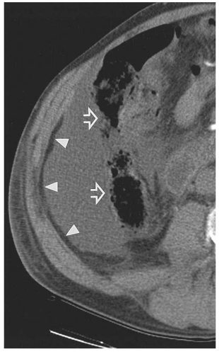
Ct Showing Free Fluid In The Right Paracolic Gutter No Free Air And Download Scientific Diagram

Ct Showing Free Fluid In The Right Paracolic Gutter No Free Air And Download Scientific Diagram

Http Pdf Posterng Netkey At Download Index Php Module Get Pdf By Id Poster Id 114921

Http Pdf Posterng Netkey At Download Index Php Module Get Pdf By Id Poster Id 114921

Peritoneal Cavity Anatomy In Ct Peritoneography A Comprehensive Description Semantic Scholar

Peritoneal Cavity Anatomy In Ct Peritoneography A Comprehensive Description Semantic Scholar

Paracolic Gutter

Paracolic Gutter

Paracolic Gutter

As the fluid continues superiorly into the inferior extension of the splenorenal recess and perhaps to some degree medially within the unusual variant of a retropancreatic recess.

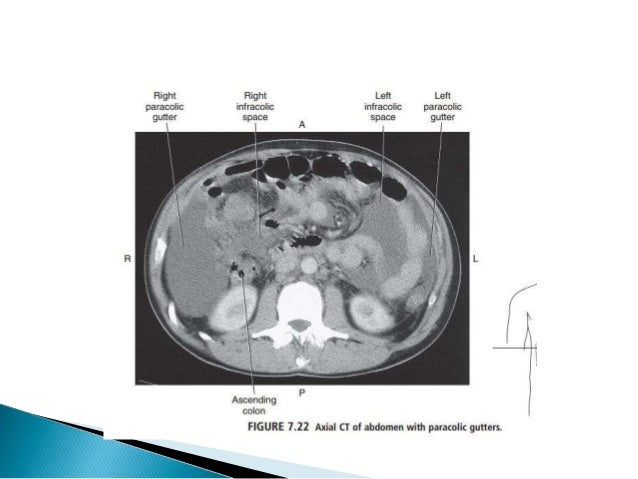
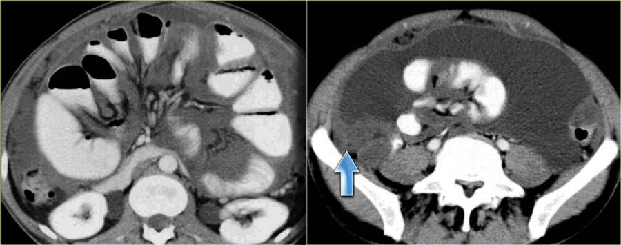
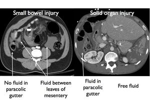
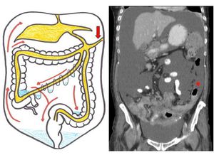
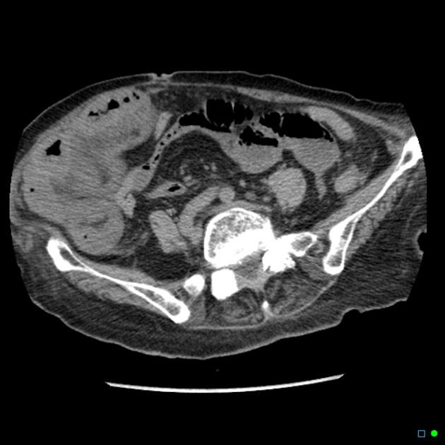
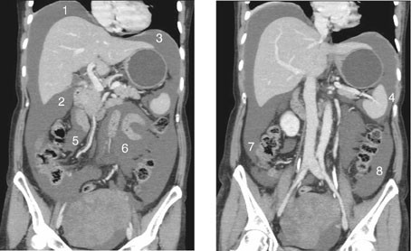
Paracolic gutter ct anatomy.

On the right hand side the right lateral paracolic gutter is found lateral to the ascending colon and medial to the lateral part of the anterior abdominal wall. The phrenicocolic ligament separates the left paracolic gutter from the left supramesocolic space it is continuous with the peritoneum of the left lateral aspect of the transverse mesocolon and the splenorenal ligament adjacent the splenic hilum 1 it attaches to the parietal peritoneum along the posterolateral aspect of the diaphragm at the level of the eleventh rib. A less obvious medial paracolic gutter may be formed especially on the right side if the colon possesses a short mesentery for part of its length. The right paracolic gutter is larger than the left and communicates freely with the right subphrenic space.

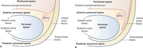
It is larger than the left paracolic gutter which is partially separated from the left subphrenic spaces by the phrenicocolic ligament. The left medial paracolic gutter. The right lateral paracolic gutter. Rarely the appearance of thickened fascia may be simulated on ct supine scans by intraperitoneal fluid within posterior peritoneal recesses particularly on the left 17 52 53 fluid layering in a deep left paracolic gutter may mimic thickening of the lateroconal fascia and a portion of the posterior renal fascia.

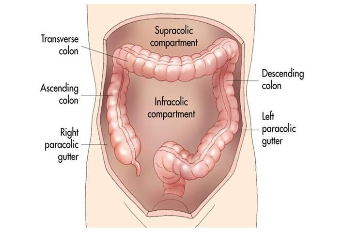
There are two paracolic gutters in the body the right and left lateral paracolic gutter. The left lateral paracolic gutter. The paracolic spaces gutters are located lateral to the peritoneal reflections of the left and right sides of the colon fig 8a. Paracolic gutters are open areas between the wall of the abdomen and the colon.

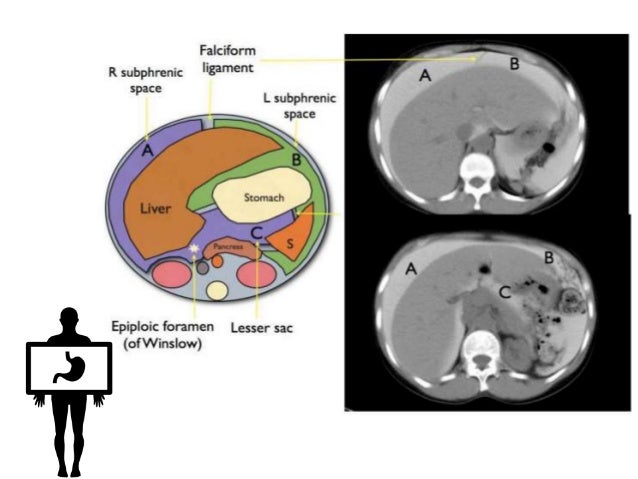
These gutters are used to drain infectious material away from the essential internal organs. Right paracolic gutter there are paracolic gutters adjacent to the infracolic compartments that are clinically significant. Both paracolic spaces are in continuity with the pelvic peritoneal spaces. The right paracolic gutter is a component of the right inframesocolic space continuous superiorly with the right subhepatic and right subphrenic spaces.

Etiologically it means a channel adjacent to the abdominal wall. It is the depression between the postero lateral wall of the abdomen and the lateral margins of the ascending and descending colon. There are two paracolic gutters. It is also known as sulci paracolic and paracolic recesses.

The paracolic gutters paracolic sulci paracolic recesses are spaces between the colon and the abdominal wall. Root of the sigmoid mesentery. Between the outer wall of the colon and back side of the abdominal wall there is an open space known as the paracolic gutter. The right and left paracolic gutters are peritoneal recesses on the posterior abdominal wall lying alongside the ascending and descending colon.

It predominantly flows up the right paracolic gutter which is deeper and wider than the left and is partially cleared by the subphrenic lymphatics.

Imaging Anatomy Of Peritoneum

Imaging Anatomy Of Peritoneum

Peritoneum Intraperitoneal Spaces

Peritoneum Intraperitoneal Spaces

Abdomen And Pelvis Radiology Key

Abdomen And Pelvis Radiology Key

The Radiology Assistant Peritoneal Pathology

The Radiology Assistant Peritoneal Pathology

Http Pdf Posterng Netkey At Download Index Php Module Get Pdf By Id Poster Id 117837

Http Pdf Posterng Netkey At Download Index Php Module Get Pdf By Id Poster Id 117837

Gastrointestinal Radiology

Gastrointestinal Radiology

Left Paracolic Gutter

Left Paracolic Gutter

Http Pdf Posterng Netkey At Download Index Php Module Get Pdf By Id Poster Id 127176

Http Pdf Posterng Netkey At Download Index Php Module Get Pdf By Id Poster Id 127176

Epos

Epos

Ct Showing Free Fluid In The Right Paracolic Gutter No Free Air And Download Scientific Diagram

Ct Showing Free Fluid In The Right Paracolic Gutter No Free Air And Download Scientific Diagram

Fitz Hugh Curtis Syndrome Radiology Case Radiopaedia Org

Fitz Hugh Curtis Syndrome Radiology Case Radiopaedia Org

Http Pdf Posterng Netkey At Download Index Php Module Get Pdf By Id Poster Id 101511

Http Pdf Posterng Netkey At Download Index Php Module Get Pdf By Id Poster Id 101511

The Spread And Localization Of Intraperitoneal Abscesses Dynamic Radiology

The Spread And Localization Of Intraperitoneal Abscesses Dynamic Radiology

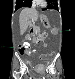
Free Intraperitoneal Fluid Summary Radiology Reference Article Radiopaedia Org

Free Intraperitoneal Fluid Summary Radiology Reference Article Radiopaedia Org

Epos

Epos

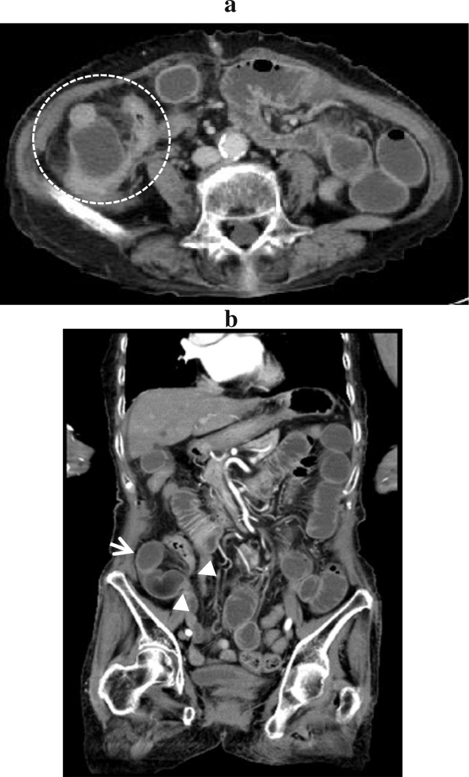
Pericaecal Internal Hernia Radiology Case Radiopaedia Org

Pericaecal Internal Hernia Radiology Case Radiopaedia Org

Pancreatic Injury Post Laparoscopic Subtotal Colectomy There Is Pancreatic Fluid Tracking From The Pancreatic Tail Arrows To Th Colectomy Abdomen Pancreatic

Pancreatic Injury Post Laparoscopic Subtotal Colectomy There Is Pancreatic Fluid Tracking From The Pancreatic Tail Arrows To Th Colectomy Abdomen Pancreatic

Peritoneal Carcinomatosis From Adenocarcinoma Of The Colon Ct Shows A Small Amount Of Ascites Nodular Soft Tissue Thick Disease Secondary Historical Figures

Peritoneal Carcinomatosis From Adenocarcinoma Of The Colon Ct Shows A Small Amount Of Ascites Nodular Soft Tissue Thick Disease Secondary Historical Figures

Https Encrypted Tbn0 Gstatic Com Images Q Tbn 3aand9gcqviiuoor7cqvjuy2tb0atddfdnjxh67jahtkkexb8 Usqp Cau

Https Encrypted Tbn0 Gstatic Com Images Q Tbn 3aand9gcqviiuoor7cqvjuy2tb0atddfdnjxh67jahtkkexb8 Usqp Cau

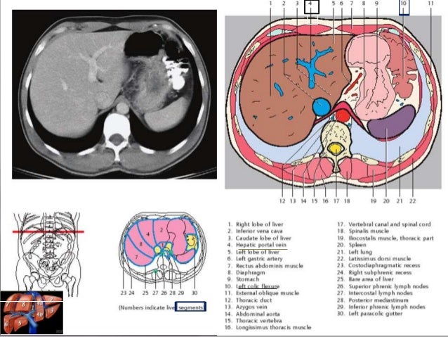
Ct Mri Upper Abdomen Normal Anatomy Dr Ahmed Eisawy Youtube

Ct Mri Upper Abdomen Normal Anatomy Dr Ahmed Eisawy Youtube

The Peritoneal Cavity Greater Sac Lesser Sac Teachmeanatomy

The Peritoneal Cavity Greater Sac Lesser Sac Teachmeanatomy

Jaypeedigital Ebook Reader

Jaypeedigital Ebook Reader

Abdomen Nontraumatic Emergencies Radiology Key

Abdomen Nontraumatic Emergencies Radiology Key

Sectional Anatomy Of Abdomen

Sectional Anatomy Of Abdomen

Epos

Epos

Epos

Epos

Figure 7 From Applied Peritoneal Anatomy Semantic Scholar

Figure 7 From Applied Peritoneal Anatomy Semantic Scholar

Paracecal Hernia Due To Membranous Adhesion Of The Omentum To The Right Paracolic Gutter Springerlink

Paracecal Hernia Due To Membranous Adhesion Of The Omentum To The Right Paracolic Gutter Springerlink

Peritoneal Collections Springerlink

Peritoneal Collections Springerlink

1 Short Descending Mesocolon And Redundant Sigmoid Colon In The Right Download Scientific Diagram

1 Short Descending Mesocolon And Redundant Sigmoid Colon In The Right Download Scientific Diagram

Pin On Abdomen

Pin On Abdomen

Computed Tomography Ct In The Evaluation Of Bladder And Ureteral Trauma Indications Technique And Diagnosis Springerlink

Computed Tomography Ct In The Evaluation Of Bladder And Ureteral Trauma Indications Technique And Diagnosis Springerlink

The Colon Ascending Transverse Descending Sigmoid Teachmeanatomy

The Colon Ascending Transverse Descending Sigmoid Teachmeanatomy

Pin By Parsa Amiri On Body Anatomy In 2020 Body Anatomy Anatomy Body

Pin By Parsa Amiri On Body Anatomy In 2020 Body Anatomy Anatomy Body

Peritoneal Cavity Vessels Nodes And Abdominal Wall Radiology Key

Peritoneal Cavity Vessels Nodes And Abdominal Wall Radiology Key

Ct Mri And Pet Imaging In Peritoneal Malignancy Abstract Europe Pmc

Ct Mri And Pet Imaging In Peritoneal Malignancy Abstract Europe Pmc

Posterior Right Subhepatic Space Radiology Reference Article Radiopaedia Org

Posterior Right Subhepatic Space Radiology Reference Article Radiopaedia Org

Cross Section At L3 4 Anatomy Round Ligament Ligamentum Teres Of Liver Transverse Colon Branches Of Inferior Epigas Anatomy Plexus Products Cross Section

Cross Section At L3 4 Anatomy Round Ligament Ligamentum Teres Of Liver Transverse Colon Branches Of Inferior Epigas Anatomy Plexus Products Cross Section

Role Of Imaging In Peritoneal Surface Malignancies Abstract Europe Pmc

Role Of Imaging In Peritoneal Surface Malignancies Abstract Europe Pmc

Renal Pelvis Radiology Reference Article Radiopaedia Org

Renal Pelvis Radiology Reference Article Radiopaedia Org

Role Of Imaging In Peritoneal Surface Malignancies Abstract Europe Pmc

Role Of Imaging In Peritoneal Surface Malignancies Abstract Europe Pmc

29 Pelvis Radiology Key

29 Pelvis Radiology Key

Patterns Of Peritoneal Spread Of Tumor In The Abdomen And Pelvis

Patterns Of Peritoneal Spread Of Tumor In The Abdomen And Pelvis

Https Encrypted Tbn0 Gstatic Com Images Q Tbn 3aand9gcsv2cwxn44g3u2 E8028wfvuyy Lqabzn8nxot9gzw Jd4hzlcd Usqp Cau

Https Encrypted Tbn0 Gstatic Com Images Q Tbn 3aand9gcsv2cwxn44g3u2 E8028wfvuyy Lqabzn8nxot9gzw Jd4hzlcd Usqp Cau

Source : pinterest.com