Orbital Roof Ct

Pin On A B I

Pin On A B I

Osteoma Of Orbital Roof Radiology Case Radiopaedia Org Radiology Radiology Imaging Diagnostic Imaging

Osteoma Of Orbital Roof Radiology Case Radiopaedia Org Radiology Radiology Imaging Diagnostic Imaging

Osteoma Of Orbital Roof Radiology Case Radiopaedia Org Radiology Brain Images Mri Brain

Osteoma Of Orbital Roof Radiology Case Radiopaedia Org Radiology Brain Images Mri Brain

Neurofibromatosis Type 1 Orbital Manifestations Radiology Case Radiopaedia Org Dysplasia Of The Neurofibromatosis Type 1 Manifestation Radiology Imaging

Neurofibromatosis Type 1 Orbital Manifestations Radiology Case Radiopaedia Org Dysplasia Of The Neurofibromatosis Type 1 Manifestation Radiology Imaging

Head Ct Scan Procedure Radtechonduty Ct Scan Brain Anatomy Radiography

Head Ct Scan Procedure Radtechonduty Ct Scan Brain Anatomy Radiography

Transitional Cell Carcinoma Tcc Is The Most Common Primary Neoplasm Of The Bladder And Bladder Tcc Is The Most Common Tumour Of The Entire Urinary Syst

Transitional Cell Carcinoma Tcc Is The Most Common Primary Neoplasm Of The Bladder And Bladder Tcc Is The Most Common Tumour Of The Entire Urinary Syst

Transitional Cell Carcinoma Tcc Is The Most Common Primary Neoplasm Of The Bladder And Bladder Tcc Is The Most Common Tumour Of The Entire Urinary Syst

It is separated posteriorly from roof of orbit by superior orbital fissure and separated from floor of orbit by inferior orbital fissure the lacrimal foramen which transmits the recurrent meningeal branch of the ophthalmic artery is located anterior to the superior orbital fissure along the superior edge of the lateral wall.

Orbital roof ct.

This frequently causes downward and forward displacement of the globe. Although sagittal sections are also helpful in some cases the axial images are less so. Most orbital roof fractures are blow in fractures displacement of the bone is towards the orbit. Preoperative ct imaging needs to be checked for unusual pneumatization of the orbital roof and possible weak spots.

The ultimate diagnosis is made by computed tomography ct of the face. While this article will try to list most of the important features of the orbital roof it is by no means comprehensive. The gold standard for diagnosis of an orbital roof fracture is thin cut coronal ct scanning of the face orbits. It is a thin lamina separating the orbit anteriorly from the frontal sinus and posteriorly from the anterior cranial fossa.

Large round bony mass protruding superior and medially from the left anterior cranial fossa floor. The orbital roof is composed of the orbital plate of the frontal bone with a small contribution from the lesser wing of the sphenoid at the apex figures 3 4 and 3 5. The orbital roof largely consists of the orbital process of the frontal bone. There are several structures and features regarding the orbital roof that we need to remember.

This fissure allows the passage to. Clinical diagnosis is based on meticulous examination of the eye including patient vision and palpation of the orbital aperture. Contrast is not needed. Mild surround frontal lobe edema.

Orbital process of the frontal bone orbital process of the zygomatic bone. Orbital process of the frontal bone anterior superior portion lesser wing of the sphenoid postero medial portion inferior wall.

Pin On Radiography

Pin On Radiography

Pin On Ct Scans

Pin On Ct Scans

Neurofibromatosis Type Ii Radiology Case Radiopaedia Org Radiology Neurology Brain Images

Neurofibromatosis Type Ii Radiology Case Radiopaedia Org Radiology Neurology Brain Images

Small Bowel Infarction Radiology Case Radiopaedia Org Radiology Bowels Ct Scan

Small Bowel Infarction Radiology Case Radiopaedia Org Radiology Bowels Ct Scan

Pin By Juliann Marie On Gu Radiology Pet Ct Radiology Medical

Pin By Juliann Marie On Gu Radiology Pet Ct Radiology Medical

Tate Of The Art Ct Scanners Are So Fast We Can Image The Coronary Arteries To Look For Atherosclerotic Plaque With Images Diagnostic Imaging Medical Imaging Ct Scan

Tate Of The Art Ct Scanners Are So Fast We Can Image The Coronary Arteries To Look For Atherosclerotic Plaque With Images Diagnostic Imaging Medical Imaging Ct Scan

Blowout Orbital Fracture Radiology Medical Imaging Radiography

Blowout Orbital Fracture Radiology Medical Imaging Radiography

Pin On Ct Scans

Pin On Ct Scans

Subdural Acute On Chronic Medical Imaging Brain Images

Subdural Acute On Chronic Medical Imaging Brain Images

Fibrous Dysplasia Frontal A And Lateral B Plain Radiographs Show A Well Defined Thickened And Sclerotic Appearanc Pediatrics Pathology

Fibrous Dysplasia Frontal A And Lateral B Plain Radiographs Show A Well Defined Thickened And Sclerotic Appearanc Pediatrics Pathology

The Racing Car Sign Refers To Widely Spaced Lateral Ventricles Due To Agenesis Of The Corpus Callosum With Intervening Prob Radiology Corpus Callosum Car Signs

The Racing Car Sign Refers To Widely Spaced Lateral Ventricles Due To Agenesis Of The Corpus Callosum With Intervening Prob Radiology Corpus Callosum Car Signs

Pleomorphic Xanthoastrocytoma Radiology Reference Article Radiopaedia Org Radiology

Pleomorphic Xanthoastrocytoma Radiology Reference Article Radiopaedia Org Radiology

Pin On Signs

Pin On Signs

Axial Ct Image In A Patient With Cholangiocarcinoma Shows Enlarged Prepancreatic Yellow And Retroperitoneal Lymph Nodes In 2020 Lymph Nodes Malignant Tumor Lymphoma

Axial Ct Image In A Patient With Cholangiocarcinoma Shows Enlarged Prepancreatic Yellow And Retroperitoneal Lymph Nodes In 2020 Lymph Nodes Malignant Tumor Lymphoma

Cerebrovascular Imaging Head And Neck Radiology Movie Posters

Cerebrovascular Imaging Head And Neck Radiology Movie Posters

Bones And Xray Photos Google Search Medical Radiography Radiology Facial Bones

Bones And Xray Photos Google Search Medical Radiography Radiology Facial Bones

Avm Ct Google Search Central Nervous System Nervous System Nervous

Avm Ct Google Search Central Nervous System Nervous System Nervous

Ct Abdomen Arabic Prof Mamdouh Mahfouz Youtube

Ct Abdomen Arabic Prof Mamdouh Mahfouz Youtube

Subependymal Giant Cell Astrocytomas But Watch Out They Are Not Astrocytomas Really Are Another Classic Tuberous Sclerosis Radiology Brain Images

Subependymal Giant Cell Astrocytomas But Watch Out They Are Not Astrocytomas Really Are Another Classic Tuberous Sclerosis Radiology Brain Images

Clear Cell Rcc Left Kidney Ctisus Com Case Study Medical Imaging Pediatrics

Clear Cell Rcc Left Kidney Ctisus Com Case Study Medical Imaging Pediatrics

Pin On Ct Scans

Pin On Ct Scans

Leg Ct Scan Shows Multiple Bullet Fragments In A Patient Who Was Shot Through The Calf Radiologist Radiology T Diagnostic Imaging Radiology Ct Scan

Leg Ct Scan Shows Multiple Bullet Fragments In A Patient Who Was Shot Through The Calf Radiologist Radiology T Diagnostic Imaging Radiology Ct Scan

Ct Of The Facial Skeleton Of A 40 Year Old Woman Demonstrates Extensive Bony Expansion With Areas Of Heterogeneous Nursing Study Nuclear Medicine Head And Neck

Ct Of The Facial Skeleton Of A 40 Year Old Woman Demonstrates Extensive Bony Expansion With Areas Of Heterogeneous Nursing Study Nuclear Medicine Head And Neck

Pin On Ct Scans

Pin On Ct Scans

Pin On Pinterest Finds

Pin On Pinterest Finds

Craniopharyngioma Radiology Pediatrics Neonatal

Craniopharyngioma Radiology Pediatrics Neonatal

Anatomy The Orbit Is A Bony Pyramid With The Optic Foramen At Its Apex Floor Of Orbit Is The Roof Of Ma Types Of Fractures Maxillary Sinus Nursing Information

Anatomy The Orbit Is A Bony Pyramid With The Optic Foramen At Its Apex Floor Of Orbit Is The Roof Of Ma Types Of Fractures Maxillary Sinus Nursing Information

Pin On Ct Scans

Pin On Ct Scans

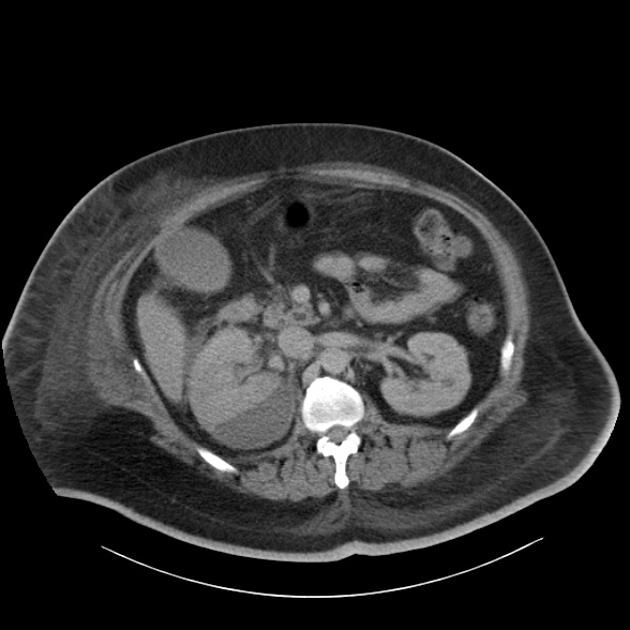
Subcapsular Renal Collection Causing A Page Kidney Radiology Case Radiopaedia Org Renal Radiology Medical Imaging

Subcapsular Renal Collection Causing A Page Kidney Radiology Case Radiopaedia Org Renal Radiology Medical Imaging

Mineralizing Angiopathy Radiology Brain Images Adolescent Health

Mineralizing Angiopathy Radiology Brain Images Adolescent Health

Pin By Pankaj Kaira On Radiology Interesting Cases And Spotters Radiology Picture Video Medical

Pin By Pankaj Kaira On Radiology Interesting Cases And Spotters Radiology Picture Video Medical

Is Case 332 Bronchial Atresia Radiology Bronchial Pet Ct

Is Case 332 Bronchial Atresia Radiology Bronchial Pet Ct

Alberta Stroke Program Early Ct Score Aspects Radiology Reference Article Radiopaedia Org

Alberta Stroke Program Early Ct Score Aspects Radiology Reference Article Radiopaedia Org

Sclerosing Encapsulating Peritonitis Radiology Reference Article Radiopaedia Org Radiology Pathology Small Bowel Obstruction

Sclerosing Encapsulating Peritonitis Radiology Reference Article Radiopaedia Org Radiology Pathology Small Bowel Obstruction

Kartegener S Syndome Dyskinetic Cilia Syndrome Upper Ct Shows Dextrocardia And Bronchiectasis At Both Bases Lower Ct Shows Situs Invers Imagenologia

Kartegener S Syndome Dyskinetic Cilia Syndrome Upper Ct Shows Dextrocardia And Bronchiectasis At Both Bases Lower Ct Shows Situs Invers Imagenologia

Right Sided Diverticulitis Ct Shows An Inflamed Cecal Diverticulum Arrowhead With Regional Colonic Wall Thickening Radiologia Imagenologia Tomografia

Right Sided Diverticulitis Ct Shows An Inflamed Cecal Diverticulum Arrowhead With Regional Colonic Wall Thickening Radiologia Imagenologia Tomografia

The Spoke Wheel Sign Refers To The Pattern Of Vessels Coursing Through Meningiomas When Seen In Cross Section It Is The Result Radiology Pet Ct White Matter

The Spoke Wheel Sign Refers To The Pattern Of Vessels Coursing Through Meningiomas When Seen In Cross Section It Is The Result Radiology Pet Ct White Matter

Archaeologists Found A Medieval Body With A Tumor That Was Growing Teeth But That S Not The Wh Archeologist Ancient Origins Historical Figures

Archaeologists Found A Medieval Body With A Tumor That Was Growing Teeth But That S Not The Wh Archeologist Ancient Origins Historical Figures

Pyelonephritis The Frequency Of Tomographic Findings For The Several Parameters Was The Following Striated Nephrogram 100 Renal Enlargement 65 Perirenal

Pyelonephritis The Frequency Of Tomographic Findings For The Several Parameters Was The Following Striated Nephrogram 100 Renal Enlargement 65 Perirenal

Radyo Panosundaki Pin

Radyo Panosundaki Pin

Pin On Pet Ct

Pin On Pet Ct

Dandy Walker Malformation Consists Of A Group Of Anomalies Where There Is A Posterior Fossa Cyst Communicates With The Radiology Pediatric Radiology Neurology

Dandy Walker Malformation Consists Of A Group Of Anomalies Where There Is A Posterior Fossa Cyst Communicates With The Radiology Pediatric Radiology Neurology

Pin On Radiologie

Pin On Radiologie

Source : pinterest.com